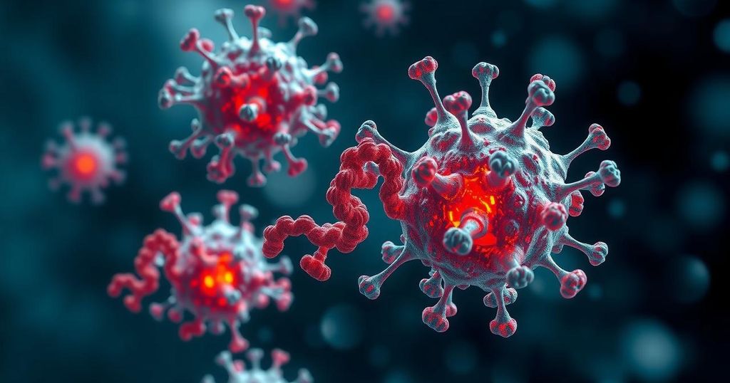
Stanford Scientists Harness Apoptosis to Flip Cancer Cells’ Fate
Stanford researchers have developed a novel technique that encourages cancer cells to self-destruct by artificially linking two proteins, BCL6 and CDK9. This method reignites apoptosis genes typically silenced in cancer, offering a more targeted cancer therapy that could avoid the collateral damage associated with traditional cancer treatments. Their findings suggest a promising future for precise and effective cancer treatment strategies.
Stanford researchers are revolutionizing cancer treatment with a groundbreaking technique that utilizes the body’s natural process of cell death, known as apoptosis, to promote the self-destruction of cancerous cells. By ingeniously gluing together two proteins—BCL6, which when mutated contributes to lymphoma, and CDK9, an enzyme that activates gene expression—they have created a compound that can switch on apoptosis genes that cancer cells typically keep off. Known as “molecular glue,” this method alters the cancer cells’ dependency on BCL6, sparking their demise instead of allowing them to thrive.
This innovative concept was birthed during a contemplative walk in the enchanting forests of Kings Mountain by Gerald Crabtree, MD, a revered cancer biologist at Stanford. Reflecting on the fundamental mechanisms of biology, Crabtree noted the significance of apoptosis, a process vital for maintaining organ health and immune balance. His inspiration to tackle cancer through this natural bottleneck was both compelling and audacious, aiming to replicate the high specificity with which the body naturally eliminates around 60 billion cells daily, without collateral damage.
The team’s journey uncovered how BCL6—a driver of diffuse large B-cell lymphoma—silences apoptosis-promoting genes, fostering cancer’s notorious immortality. By coupling BCL6 with CDK9, the researchers crafted a molecular agent capable of reigniting that silenced cellular machinery, potentially tipping the scales from survival to self-erasure. In laboratory tests, this fusion exhibited a potent lethality against the targeted cancer cells while sparing healthy ones, underscoring its hopeful implications in future therapies.
This approach contrasts sharply with traditional cancer therapies like chemotherapy, which indiscriminately damage healthy cells. The researchers believe that the dual activation of multiple death signals through their polymer invention could outwit the adaptability of cancer, preventing it from developing resistance. “It’s sort of cell death by committee,” remarked one of the postdoctoral scholars involved in the project.
As they look toward further tests in living organisms and clinical trials through their startup, Shenandoah Therapeutics, the excitement and optimism surrounding this innovative cancer therapy blend promise a new era in the fight against cancer, embracing both the wisdom of nature and the mastery of scientific ingenuity.
The study of apoptosis, or programmed cell death, is crucial in understanding how the body maintains balance and health among its cells. While traditionally viewed as a detrimental characteristic in cancer, researchers are now exploring creative ways to exploit the very mechanisms that allow cancer cells to proliferate unchecked. By utilizing understanding from cell biology and the specific role of certain proteins, scientists are pioneering therapeutics that could lead to more effective and selective cancer treatments, minimizing collateral damage to healthy cells that has long plagued conventional approaches.
The Stanford researchers’ innovative strategy to manipulate apoptosis for fighting cancer represents a novel frontier in medical science, potentially heralding a shift from merely suppressing cancer cells to actively engaging them in their demise. By focusing on the specific dependencies of cancer cells, this emerging therapy not only promises to enhance treatment efficacy but also aims to overcome resistance—creating a multi-faceted approach to tackling cancer that aligns with natural biological processes. The pathway they’ve embarked on could redefine cancer treatment protocols and inspire further research across various cancer types.
Original Source: med.stanford.edu


Post Comment